top of page
Writer's pictureDr Chandan Dolare

Maxillofacial trauma imaging using CBCT

The diagnosis of maxillofacial fractures is accomplished by a combination of clinical examination and imaging investigations. The radiographic examination of any suspected fracture should be in at least two planes, oriented perpendicular to each other.

CBCT has been used to assess maxillofacial and mandibular fractures. Limited FOV CBCT is also capable of detecting jaw and tooth fractures. CBCT is superior to conventional CT imaging modality to assess bone and tooth related structures. The examination includes orthogonal sections (axial, sagittal and coronal sections). It also includes multiplanar sections which can be reconstructed in any desired plane and 3D reconstruction, which has proved to be very valuable in the evaluation of complex maxillofacial anatomy. The 3d images help in visualization of the displaced fracture segments and their relationship to one another. This blog discusses maxillofacial and mandibular traumas and fractures from reported cases.


1. Fracture of the alveolar process (non-displaced) : Cross sectional images and coronal images at 180µm showing coronal defect approximating pulp space i.r.t 21. Fracture line is noted with the distal socket wall at the cervical 3rd level. The fractured portions are not displaced. Localized osteolytic lesion is noted extending till midroot level i.r.t 21. The lesion shows perforation of the labial cortical plate. Apical resorption is noted.


2. Nondisplaced alveolar process fracture with symphysis region : Coronal images at 300µm show a fracture line extending from the labial plate at the cervical 3rd level i.r.t 31 to 42 region involving the interdental bone. It involves the socket walls i.r.t 41,42. Minimal lingual displacement of the lingual plate is noted i.r.t distal aspect of 42. Apical hypodensity is noted with 41,42. Widening of periodontal ligament space is noted with 31.


3. Bilateral dentoalveolar fracture (displaced) with body of mandible : Coronal images at 300µm showing a fracture line with the right body of mandible extending superoinferiorly from the buccal to the lingual cortical plate in a lateromedial direction. The fracture line runs from the mesial interdental bone i.r.t 47 to the inferior border of mandible at the right angle of mandible. The socket of 47 is involved. The inferior alveolar canal is involved and the fractured segment is displaced laterally.

The 2nd fracture defect involves the distal interdental bone i.r.t 38. The defect involves the buccal and lingual cortices and runs posteroinferiorly from the socket i.r.t 38 to the inferior border at the left angle of mandible. The inferior alveolar canal is involved. The fractured segment is displaced mesially.


4. Multiple, displaced, fractures involving the maxillofacial complex and mandible :

3d images and axial sections at 300µm showing fracture defect with the lateral wall of the right orbital cavity.

Displaced fracture is noted with the left zygomaticomaxillary complex along with dentoalveolar fracture i.r.t 24-27 region. Fracture involves the left orbital floor. Fracture noted with the left zygomatic arch. Potential left intrasinus hemorrhage noted within the left maxillary sinus. Bilateral fracture noted with the frontal process of maxilla.

Displaced dentoalveolar fracture is noted with the mandible i.r.t 44-36 region. Vertical impaction is noted with 33,43 within the symphysis of mandible.

Coronal sections show bilateral fracture with the frontal process of maxillae or nasal process fracture.


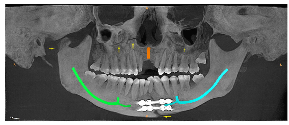
5. Displaced fracture of the maxilla, anterior mandible and right condylar process :

Panoramic image, cross sectional images and axial sections at 300µm show dentoalveolar fracture with the right anterior maxilla i.r.t 13,14 region. Potential surgical defect or fracture defect is noted with the lateral wall of the left maxillary sinus showing approximation with the left zygoma. Fracture is noted with the neck of the right condylar process, which is displaced anteriomedially in an inferior direction.

Partially healed and misaligned dentoalveolar fracture is noted i.r.t 32-41 region with the symphysis region of mandible.

Enamel-dentin-pulp fracture and apical abscess noted with 12,22.


6. Dentoalveolar fracture and intrusion injury with anterior maxilla in a pediatric patient : Cross sectional and coronal sections at 90µm show intrusion injury with 11. Intrusion of 21 is noted within the nasal fossa. Apices of 21 project into the left inferior meatus. Potential concussion injury noted i.r.t 12,22. Crown fracture and dentoalveolar fracture is noted with 53. Suspected discontinuity is noted with the buccal cortical plate i.r.t 55 region. Bilateral, localized, mild, maxillary sinus mucositis noted.


7. Non-displaced fracture with the left condylar head : Anteroposterior sections with the left TMJ at 300µm show fracture line involving the left condylar head. Fracture line is noted involving the articular surface and the lower portion of the condylar head extending from the mesial to the distal aspect. Separation is noted along with a step defect along the lateral aspect and the distal aspect of the condylar head.

Coronal sections at 300µm show fracture with the right and left nasal bone. Horizontal crown fracture involving pulp space is noted i.r.t 14. Crown fracture is noted with 46 extending into buccal socket wall. Crown fracture involving pulp space and apical periodontitis noted with 15. Vertical fracture is noted involving coronal pulp space i.r.t 16. Horizontal root fracture and vertical crown fracture is noted with 24. Vertical crown fracture involving coronal pulp space noted with 25. Fracture involving distal aspect of the crown noted with 26. Fracture involving distobuccal cusp noted with 27. Multiple coronal fractures noted with 35. Crown fracture involving pulp space noted with 36. Mesiolingual cusp fracture noted with 38.


8. Bilateral zygomatic arch fracture and dentoalveolar fractures : Axial and coronal sections at 300µm show fracture line/s with the lateral wall of the right and left orbital cavities. Discontinuity is noted with the left nasal bone.

Bilateral, displaced zygomatic arch fracture is noted. Displaced left condylar neck fracture is noted. Displaced dentoalveolar fracture is noted with the symphysis region of mandible. Interdental wiring noted i.r.t 31-32 region. Enamel and dentin fracture noted i.r.t 28,38.


9. Bilateral fracture noted with the right and left naso-orbito-ethmoid complex :

Coronal and axial images at 200 µm show fracture involving the right and left naso-orbito-ethmoid complex. Opacification is noted with the right and left ethmoidal and left frontal air sinuses suggestive of intrasinus hematoma. Suspected dentoalveolar fracture is noted with 15 region. Fracture is noted with the right and left maxillary sinuses. Bilateral, localized radiodensities noted within the maxillary sinuses suggestive of intrasinus hematoma or localized mucositis.


References :

Oral Radiology : Principles and interpretation 5th edition White & Pharoah

516 views1 comment

1 Comment


shikhsmumbai
Sep 15, 2020

Very well explained Dr chandan...This can simplify our radiographic diagnosis

Like
logo use.jpg
bottom of page